Disclaimer: Early release articles are not considered as final versions. Any changes will be reflected in the online version in the month the article is officially released.
Author affiliation: Unité Mixte de Recherche de l’Institut National de Recherche pour l’Agriculture, l’Alimentation, et l’Environnement 1225 Interactions Hôtes-Agents Pathogènes, École Nationale Vétérinaire de Toulouse, Toulouse, France (T. Barrio, J.-Y. Douet, A. Huor, S. Lugan, N. Aron, H. Cassard, O. Andréoletti); Norwegian Veterinary Institute, Ås, Norway (S.L. Benestad); Consejo Superior de Investigaciones Científicas, Madrid, Spain (J.C. Espinosa, J.M. Torres); Universidad de Zaragoza, Zaragoza, Spain (A. Otero, R. Bolea)
Chronic wasting disease (CWD) is a highly contagious prion disease affecting members of the Cervidae family. CWD is widely spread across North America, where it endangers the survival of free-ranging cervid populations. In Europe, CWD was reported in a reindeer (Rangifer tarandus tarandus) from Norway in 2016 (1). Since 2016, several cases have been reported in Norway, Sweden, and Finland in multiple species, including reindeer, red deer (Cervus elaphus), and moose (Alces alces) (2).
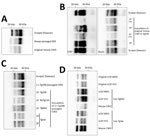
Whereas CWD strains circulating in North America exhibit some uniformity (3), the cases found in Europe are more variable. Transmission into rodent models has revealed multiple CWD strains that are apparently different than strains in North America, and moose cases in Norway have demonstrated biochemical patterns distinct from previous cases in Europe (4). We characterized the interspecies transmission potential of 1 moose CWD isolate from Norway (Norwegian Veterinary Institute identification no. 16–60-P153) (4) by intracerebral injection of mouse models expressing the normal prion protein (PrPC) sequences from several species (Figure, panel A).
We anesthetized and inoculated 6–10-week-old mice with 2 mg of equivalent tissue (20 µL of 10% brain homogenate) in the right parietal lobe. We monitored the inoculated animals daily and humanely euthanized animals at the onset of clinical signs or after the preestablished endpoint of 700 days postinfection (dpi). We conducted a systematic proteinase K–resistant prion protein (PrPres) detection by using Western blot.
Inoculation of the original CWD isolate did not cause the propagation of detectable prions in Tg340 mice expressing methionine (TgMet) or Tg361 mice expressing valine (TgVal) at position 129 of human PrPC. We did not observe PrPres in brain tissue or disease occurrence in bovine PrPC-expressing mice (BoTg110) after intracerebral inoculation of the CWD isolate (Table; Figure, panel B).
We inoculated the CWD isolate in Tg338 mice, which overexpress ovine PrP ≈8 times. At 612 and 717 dpi (Table), 2 of 12 animals showed clinical signs of prion disease and we detected PrPres accumulation in their brain tissue (Figure, panel B). Of note, the 2 animals showed different PrPres banding patterns, with the nonglycosylated band migrating to 19 kDa in the first mouse and to 21 kDa in the second. Both PrPres-containing brains transmitted disease with 100% efficacy to second-passage Tg338 mice, which contained 21-kDa PrPres in their brains (Figure, panel B). A third passage resulted in the incubation period shortening (95 ± 5 dpi). Our observations are consistent with a progressive adaptation of the moose CWD prion to the ovine-PrPC expressing model and suggest moose CWD prions in Europe may have an intrinsic capability to propagate in ovine species with the VRQ genotype.
We next determined whether adaptation of this moose CWD agent to Tg338 altered its capacity to cross species barriers. For that purpose, we inoculated Tg338-adapted moose CWD prions (passaged twice in Tg338) to the same panel of PrPC-expressing mice models. Inoculation of the Tg338-adapted isolate to BoTg110 resulted in 100% disease transmission that showed a banding pattern and intermediate molecular weight from 19–21 kDa (Figure, panel C; Appendix Figure) and an incubation period of 431 ± 32 dpi (Table), which suggested the lack of a major transmission barrier. In addition, 1 of 8 inoculated TgMet mice showed clinical signs at 561 dpi (Table). PrPres in the brain of that mouse was revealed by a mixed 19 + 21 kDa banding pattern (Figure, panel C). A second passage in TgMet is underway.
Inoculation of TgVal resulted in efficient transmission (5/6 animals); the mean incubation period was 483 ± 35 dpi (Table) and accumulation was 21 kDa PrPres (Figure, panel C). On second passage, transmission was 100% and we observed a shorter incubation period (311 ± 12 dpi).
The incubation periods and PrPres biochemical profiles of the CWD prions that propagated in the TgMet and TgVal mice greatly differed from those observed in mice inoculated with the most prevalent human prion strains or with classic bovine spongiform encephalopathy (BSE), sheep-adapted BSE, or Tg338-adapted c-BSE (Table; Figure, panel D). Those results might suggest this CWD-derived prion strain differs from other strains documented in those models. Further investigation is necessary.
The evolution of moose CWD zoonotic potential after its passage in an ovine PrPC-expressing host is reminiscent of the well-documented altered capacities of the c-BSE agent to cross the human species barrier after adaptation in sheep and goats (9). The codon 129-dependent response to infection of humanized mice with Tg338-adapted CWD is also compatible with studies demonstrating the role of this polymorphism in susceptibility to prions (10).
In summary, our results demonstrate the potential capacity of some CWD agents to transmit to sheep or other farmed animals. Our results highlight the need to experimentally assess and monitor this transmission risk under natural exposure conditions. In addition, the dramatic changes of the zoonotic capacity of the CWD isolate we documented from Europe clearly demonstrate the risk adaptation and propagation of cervid prions into farmed animals represents. Although additional studies are needed to characterize these emerging agents, our findings have major potential implications for animal and public health.
Dr. Barrio is a research scientist with Institut National de Recherche pour l’Agriculture, l’Alimentation et l’Environnement. His research interests include animal and human prion diseases and other neurodegenerative disorders linked to misfolded proteins.
